Auswirkungen der bilateralen Diskektomie und bilateralen Diskopexie auf die Ruminationskinematik von schwarzen Merino-Schafen: TEMPOJIMS — Phase 1 — pilotierte, verblindete, randomisierte präklinische Studie
Maschinenübersetzung
Der Originalartikel ist in EN Sprache (Link zum Lesen) geschrieben.
Zusammenfassung
Hintergrund: Die Studie zur Interposition des Kiefergelenks (TEMPOJIMS) ist eine rigorose präklinische Studie, die in 2 Phasen unterteilt ist. In Phase 1 untersuchten die Autoren die Rolle des Kiefergelenkdisks und in Phase 2 bewerteten die Autoren 3 verschiedene Interpositionsmaterialien. Die vorliegende Arbeit von TEMPOJIMS - Phase 1, untersuchte die Auswirkungen der bilateralen Diskektomie und Diskopexie auf das Kauen und Wiederkäuen bei Schafen.
Methoden: Diese randomisierte, verblindete und kontrollierte präklinische Studie (in Übereinstimmung mit den ARRIVE-Richtlinien) wurde an 9 schwarzen Merinoschafen durchgeführt, um Veränderungen im Kauen und Wiederkäuen nach bilateraler Diskektomie und bilateraler Diskopexie zu bewerten, indem sie mit einer Kontrollgruppe nach Scheinoperation verglichen wurden. Die bewerteten Ergebnisse waren: (1) absolute Kauzeit; (2) Wiederkäuzeit pro Zyklus; (3) Wiederkäu-Kinematik und (4) Wiederkäu-Fläche. Nach der Baseline-Bewertung und den chirurgischen Eingriffen wurden die Ergebnisse über 3 aufeinanderfolgende Tage, alle 30 Tage, über einen Zeitraum von 6 Monaten aufgezeichnet.
Ergebnisse: Der erste Monat nach der Intervention schien der kritische Zeitraum für signifikante kinematische Veränderungen in den Gruppen mit Diskektomie und Diskopexie zu sein. Allerdings wurden 6 Monate nach den bilateralen Interventionen keine signifikanten Veränderungen im Vergleich zur Kontrollgruppe festgestellt.
Schlussfolgerungen: In dieser Studie hatten bilaterale Diskektomie und Diskopexie keinen signifikanten Einfluss auf das Kauen und die Wiederkäubewegung. Die Einführung der kinematischen Bewertung stellt eine neue Herausforderung dar, die zur Verbesserung zukünftiger Studien im Bereich des Kiefergelenks beitragen könnte.
Einleitung
Der Bereich der Bioengineering des Kiefergelenks (TMJ) wächst schnell, und das Potenzial zur Entwicklung eines interpositionalen Disc für das TMJ ist enorm. Daher sind strenge präklinische Studien erforderlich für den normalen Fortschritt der translationalen Medizin. Bevor jedoch wertvolle Ressourcen und Mittel für das TMJ-Bioengineering eingesetzt werden, ist es wichtig, unser Verständnis der durch gängige chirurgische Interventionen am Kiefergelenk induzierten Effekte zu verbessern.
Die TMJ-Diskektomie ist die am häufigsten durchgeführte intrakapsuläre Operation. Mit insgesamt guten Ergebnissen bleibt diese Technik eine vernünftige Wahl für interne Störungen, die nicht auf nicht-chirurgische Behandlungen ansprechen (Nyberg et al., 2004; Eriksson und Westesson, 2001; Mazzonetto und Spagnoli, 2001; Bjørnland und Larheim, 2003; Trumpy und Lyberg, 1995). Dennoch ist es eine umstrittene Technik, da sie die strukturellen oder biologischen Eigenschaften des TMJ nicht wiederherstellt (Takaku et al., 2000). Die TMJ-Diskopexie ist eine weniger invasive Operationstechnik, die verwendet wird, um die ideale Position des TMJ-Diskus wiederherzustellen, jedoch mit variablen Ergebnissen (Sharma et al., 2010).
Trotz der großen Anzahl an jährlich durchgeführten Diskektomie- und Diskopexie-Verfahren gab es nach unserem besten Wissen keine randomisierten, verblindeten, kontrollierten Studien, die die Auswirkungen der Kieferbewegung bei bilateraler Diskektomie und bilateraler Diskopexie an Menschen oder Tieren untersucht haben.
Klein-, mittel- und großtierische Modelle wurden verwendet, um die histologischen Effekte der einseitigen Diskektomie zu untersuchen (Bjørnland und Haanaes, 1999; Dimitroulis und Slavin, 2006; Ogi et al., 1999; Sato et al., 2002; Tong und Tideman, 2000), was zu unterschiedlichen Ergebnissen führte, von geringfügigen degenerativen Veränderungen bis hin zu TMJ-Angylosen. Diese heterogenen Ergebnisse sind wahrscheinlich auf Einschränkungen hinsichtlich der Tierwahl, des Studiendesigns und der Verwendung eines einseitigen Ansatzes mit der kontralateralen Seite als Kontrolle zurückzuführen, was möglicherweise zu Verzerrungen in den verfügbaren Ergebnissen geführt hat (Cohen et al., 2014).
Wie in einer Umfrage berichtet, die vom National Centre for the Replacement, Refinement and Reduction of Animals in Research (NC3Rs) in Auftrag gegeben wurde (Kilkenny et al., 2009), gaben nur 59% der 271 zufällig ausgewählten Artikel an, die Hypothese oder das Ziel der Studie sowie die Anzahl und Merkmale der verwendeten Tiere (d.h. Art/Stamm, Geschlecht und Alter/Gewicht) an. Die meisten der befragten Arbeiten berichteten nicht über die Verwendung von Randomisierung (87%) oder Verblindung (86%), um Verzerrungen bei der Tierauswahl und der Ergebniseinschätzung zu reduzieren. Nur 70% der Publikationen, die statistische Methoden verwendeten, beschrieben diese vollständig und präsentierten die Ergebnisse mit einem Maß für Präzision oder Variabilität (Kilkenny et al., 2009). Diese Ergebnisse sind besorgniserregend und stimmen mit Übersichten über viele Forschungsbereiche, einschließlich klinischer Studien, überein, die in den letzten Jahren veröffentlicht wurden (Kilkenny et al., 2009; Sharma et al., 2010; Van der Worp et al., 2010). Darüber hinaus haben sich die meisten früheren Studien auf histologische und bildgebende Unterschiede konzentriert, aber zusätzliche Eingaben sind entscheidend, um ein klares Verständnis der TMJ-Funktionalität zu erhalten.
In diesem Papier berichten die Autoren erstmals über eine hochwertige, präklinische Studie, die die Auswirkungen der bilateralen Diskektomie und der bilateralen Diskopexie auf die Kinematik des Kauens und Wiederkäuens bei schwarzen Merino-Schafen im Vergleich zu einer Kontrollgruppe mit Scheinoperation bewertet.
Die Bewertung der Kinematik des Kauens und Wiederkäuens des Schafskiefers basierte auf den normalen Prozessen, die von Wiederkäuern verwendet werden, um partikuläre Trockenmasse abzubauen: (1) anfängliches Kauen während des Essens und (2) weiteres Kauen während des Wiederkäuens (Pearce, 1967). Die Autoren unterschieden zwischen den beiden Prozessen und analysierten sie separat. Um das anfängliche Kauen zu analysieren, untersuchten die Autoren die Zeit, die benötigt wurde, um eine Dosis trockener Pellets zu fressen, und bezeichneten dieses Ergebnis als absolute Kaudauer. Mit diesem Ergebnis erwarteten die Autoren festzustellen, ob chirurgische Eingriffe am Kiefergelenk signifikante Veränderungen in der anfänglichen Kautime hervorrufen könnten. Um die Kauffase des Wiederkäuers zu analysieren, wurde ein spezieller Käfig geschaffen und 15 Wiederkäuzyklen mit einer Videokamera aufgezeichnet. Mit der Software Foundry Nuke (2D-Tracking) und Image J wurden die Bewegungen des Wiederkäuers in der Frontalebene analysiert, um zu erhalten: (1) Wiederkäuzeit pro Zyklus, (2) Kinematik des Wiederkäuens und (3) Wiederkäuareal.
Die Studie zum interpositionalen Material des Kiefergelenks (TEMPOJIMS) wurde mit einem strengen Design geplant, das den ARRIVE-Richtlinien entspricht (Kilkenny et al., 2009). Eine randomisierte, präklinische Studie mit verblindeten Ergebnissen war in diesem Bereich erforderlich, um die Qualität weiterer Studien zum Kiefergelenk zu erhöhen, zukünftige Behandlungsoptionen für Patienten, die sich einer Operation zum Austausch des Kiefergelenkscheibens unterziehen, zu verbessern und die Interpretation zukünftiger Studien zu interpositionalen Materialien des Kiefergelenks unter Verwendung des TEMPOJIMS-Designs zu erleichtern.
Materialien und Methoden
Die TEMPOJIMS-Studie war eine präklinische Studie, die in zwei Phasen unterteilt war (Ângelo et al., 2017). Dieses Papier konzentriert sich auf die kinematischen Ergebnisse der Phase 1, mit dem Ziel, die Auswirkungen der bilateralen Diskektomie des Kiefergelenks im Vergleich zur bilateralen Diskopexie des Kiefergelenks, im Vergleich zu einer Kontrollgruppe mit Scheinoperation, bei schwarzen Merino-Schafen zu verstehen.
Studiendesign
Die Begründung und das Protokoll für die TEMPOJIMS präklinische Studie sind öffentlich verfügbar (Ângelo et al., 2017).
Studienpopulation und Stichprobe
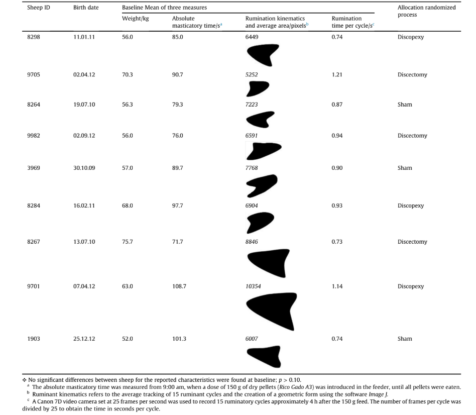
Eine Vielzahl von Stämmen/Rassen von Schafen wurde in früheren TMJ-Untersuchungen verwendet. Um die biologische Variabilität zu reduzieren, führten die Autoren diese Studie an einem schwarzen Merino-Schafstamm durch (Angelo et al., 2016). In Phase 1 verwendeten die Autoren 10 schwarze Merino-Schafe mit den folgenden Einschlusskriterien: zertifizierte schwarze Merino-Schafe, Erwachsene (im Alter von 2 bis 5 Jahren), weiblich, guter Gesundheitszustand (es wurden veterinärmedizinische Kontrollen an allen Tieren durchgeführt) und normale Zahnstellung (mit 32 Zähnen, 8 mandibulären Schneidezähnen, 12 Prämolaren und 12 Molaren).
Randomisierung
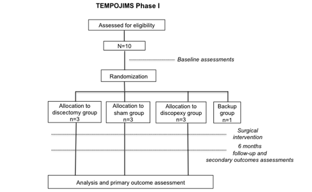
Der Randomisierungsprozess wurde von einem statistischen Team durchgeführt, das nicht an den Ergebniseinschätzungen beteiligt war. Zehn Schafe wurden zufällig den Interventionsgruppen wie folgt zugeteilt: bilaterale Diskektomie-Gruppe (n = 3), bilaterale Diskopexie-Gruppe (n = 3), Scheinoperation-Gruppe (n = 3) und Backup-Gruppe (n = 1). Das eine Backup-Schaf war für den Fall vorgesehen, dass es aufgrund von Anästhesie oder anderen nicht mit dem chirurgischen Eingriff verbundenen Komplikationen stirbt. Die Zuteilung zu jeder randomisierten Gruppe erfolgte präoperativ unter Verwendung versiegelter Umschläge (Abb. 1).
Verfahren
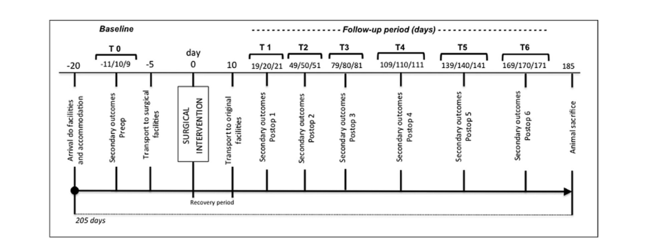
Zehn geeignete Schafe wurden ihren Basis-Pilotsekundärergebnissen zugewiesen, die an den Tagen 11, 10 und 9 vor der Operation in den TEMPOJIMS-Einrichtungen gemessen wurden (Abb. 2). Der Transport zu den chirurgischen Einrichtungen erfolgte 5 Tage vor der Operation, um Stress bei den Tieren zu vermeiden und eine Gewöhnung an die vorübergehende Unterbringung zu ermöglichen. Das chirurgische Team war nicht blind gegenüber der Behandlungszuweisung, angesichts der Art der Intervention; jedoch waren die Mitglieder des chirurgischen Teams nicht an der Bewertung der Ergebnisse beteiligt. Schwere unerwünschte Ereignisse wurden definiert als Ereignisse, die tödlich waren oder zu lebensbedrohlichen oder dauerhaften Behinderungen, mehr als 10% Gewichtsverlust pro Woche oder klinisch signifikanten Gefahren/Schäden für das Tier führten.
Anästhesieprotokoll
Fasten und Wasserrestriktion waren 24 Stunden vor der Operation erforderlich. Die Sedierung wurde mit Diazepam (0,5 mg/kg IV) durchgeführt, gefolgt von der Anästhesieeinleitung mit Ketamin (5 mg/kg IV). Eine orale Intubation wurde durchgeführt und die Anästhesie wurde mit Isofluran (1,5e2%) aufrechterhalten. Um die Analgesie des Tieres sicherzustellen, wurde am Operationstag und in den folgenden 4 Tagen Meloxicam (0,5 mg/kg IV/bid) verabreicht. Eine antibiotische Prophylaxe mit Amoxicillin und Clavulansäure wurde für 5 Tage verabreicht.
Chirurgischer Eingriff
(A) Bilateral Discektomiegruppe (n = 3): während der Allgemeinanästhesie führte das chirurgische Team einen präaurikulären Schnitt und eine stumpfe Dissektion des Weichgewebes durch, das das Gelenk bedeckt. Der Gelenkbereich wurde freigelegt und die Gelenkkapsel wurde eingeschnitten. Die Bandscheibe und ihre Befestigungen wurden identifiziert. Die medialen, anterioren, posterioren und lateralen Bandscheibenbefestigungen wurden gelöst und eine Discektomie wurde durchgeführt. Die Wunde wurde schichtweise mit Vicryl 3/0 verschlossen.
(B) Bilaterale Discopexie-Gruppe (n = 3): Während der Allgemeinanästhesie führte das chirurgische Team einen präaurikulären Schnitt und eine stumpfe Dissektion des Weichgewebes durch, das das Gelenk bedeckt. Der Gelenkbereich wurde freigelegt und die Gelenkkapsel wurde eingeschnitten. Der Diskus und seine Befestigungen wurden identifiziert. Die lateralen und hinteren Diskusbefestigungen wurden abgetrennt und mit PDS 3/0 genäht. Die Wunde wurde schichtweise mit Vicryl 3/0 verschlossen.
(C) Scheinoperation-Gruppe (n = 3): Während der Allgemeinanästhesie führte das chirurgische Team einen präaurikulären Schnitt und eine stumpfe Dissektion des Weichgewebes durch, das das Gelenk bedeckt. Die Gelenkkapsel des Kiefergelenks wurde nicht eingeschnitten. Die Wunde wurde schichtweise mit Vicryl 3/0 verschlossen.
Nachuntersuchungen
Die Baseline-Bewertung (T0) wurde vor der Operation an den Tagen —11, —10 und —9 durchgeführt (Tabelle 1). Zehn Tage nach der Operation wurden die Tiere in die TEMPOJIMS-Einrichtungen transportiert. Die Nachverfolgung der Ergebnisse begann an den Tagen 19, 20 und 21 nach der Operation (T1) und wurde alle 30 Tage über einen Zeitraum von 6 Monaten wiederholt (Abb. 2). T0eT6 basierten auf den Mittelwerten der drei Messungen. Die Bewertungen wurden von zwei speziell geschulten Gutachtern durchgeführt, die nicht mit den Interventionen verbunden waren. Alle Tiere hatten bilaterale Narben, um mögliche Verzerrungen zu reduzieren.
Kinematische Ergebnisse
Bewertete kinematische Ergebnisse waren: (1) absolute Kaudauer; (2) Wiederkäuzeit pro Zyklus; (3) wiederkäuende Kinematik; und (4) wiederkäuende Fläche.
Um die genannten Ergebnisse zu messen, wurde ein spezieller Käfig mit einem Frontfenster und einem Futterspender gebaut. Alle Bewertungen wurden von Forschern durchgeführt, die über den chirurgischen Eingriff im Unklaren waren, und waren darauf ausgelegt, Veränderungen der Kaudauer und der wiederkäuenden Kinematik zu bewerten. Diese Ergebnisse waren wie folgt:
- Absolute Kaudauer: basierend auf dem Bewertungszeitplan (Abb. 2), wurden um 9:00 Uhr die 10 Schafe in ihre einzelnen Käfige gesetzt. Eine Dosis von 150 g trockenen Pellets (Rico Gado A3) wurde im Futterspender eingeführt und die Zeit, die benötigt wurde, um alle Pellets zu fressen, wurde mit einem Chronometer gemessen.
- Ruminationszeit pro Zyklus: basierend auf dem Bewertungszeitplan (Abb. 2), haben wir 15 Ruminationszyklen ungefähr 4 Stunden nach dem 150 g Futter aufgezeichnet. Wir verwendeten eine Canon 7D Videokamera, um Bilder mit 25 Bildern pro Sekunde aufzunehmen. Die Anzahl der Bilder pro Zyklus wurde dann durch 25 geteilt, um die Zeit in Sekunden pro Zyklus zu erhalten.
- Ruminationskinematik: wir verwendeten die Software Foundry Nuke (2D-Tracking), um Kieferbewegungen zu verfolgen und den durchschnittlichen Ruminationszyklus zu berechnen. Mit der Software After Effects haben wir das 2D-Tracking in eine geometrische Form umgewandelt.
- Ruminationsfläche: wir bestimmten einen Durchschnitt für 15 Zyklen und erstellten eine geometrische Darstellung. Mit der Software Image J führten wir eine quantitative Messung in Pixeln der durchschnittlichen Ruminationsfläche durch.
Statistische Analyse
Die TEMPOJIMS Phase 1 randomisierte, kontrollierte, präklinische Studie verwendete zehn schwarze Merinoschafe mit einer Nachbeobachtungszeit von 6 Monaten. Die primäre Analyse testete die Auswirkungen der unabhängigen Variablen (IV)
für drei experimentelle Bedingungen: 1 = bilaterale Diskektomie; 2 = bilaterale Diskopexie; 3 = Scheinoperation, unter Verwendung einer Reihe von Pre-Tests (T0) und Post-Tests (T1 bis T6). Die abhängigen Variablen (Ergebnis
maße) waren: die Zeit, um 150 g Pellets zu fressen; die Wiederkäuzeit pro Zyklus; und die Wiederkäufläche. Diese Ereignisse wurden dreimal in den Pre-Tests gemessen, um die Invarianz der Ergebnismaße vor der chirurgischen Intervention (IV) zu fördern. Die sekundären Tests (Post-Tests) analysierten die Ergebnisse unter Verwendung von drei Messungen zu sechs Zeitintervallen (T1 bis T6), am selben Ort und zur selben Stunde wie die Pre-Tests (Abb. 2).
Alle statistischen Analysen wurden mit dem Statistical Package for Social Sciences (IBM SPSS, Version 22.0) durchgeführt. Shapiro-Wilk-Tests wurden für Pre-Tests (T0) und Post-Tests (T1 bis T6) durchgeführt, die eine normale Verteilung in allen Gruppen für alle Tests zeigten (p > 0.05), mit Ausnahme der T4- und T6-Wiederkäuflächen für Diskopexie (Shapiro-Wilk = 0.761 und 0.384; p < 0.05).
Zusätzlich wurden Levene-Statistiken durchgeführt, um die Homogenität der Varianzen zu testen. Statistisch signifikante Ergebnisse wurden bei T1, T2 und T5 für den Ruminationsbereich gefunden (Levene-Statistiken = 8.59, 6.35 und 7.82; p < 0.05), was dazu führte, dass für diese Zeitpunkte nicht-parametrische Tests berechnet wurden. Für die Pre-Tests und andere Zeitgruppen waren die Varianzen homogen (p > 0.07), was zu parametrischen Tests führte.
Eine einfaktorielle Varianzanalyse (ANOVA) (oder das nicht-parametrische Äquivalent Kruskale-Wallis-Test) wurde für die Querschnittsanalyse durchgeführt, um die Ergebnisvariablen auf den drei Ebenen der UV vor und nach der zufälligen Zuweisung zur Behandlungsgruppe zu vergleichen. Fisher LSD- und Games-Howell-Post-hoc-Tests wurden für gleich angenommenen und nicht angenommenen Varianzen durchgeführt. Für die longitudinale Analyse war der Mauchly-Test auf Sphärizität für die absolute Kaudauer nicht signifikant (Mauchly's W = 0.004; p = 0.589), was einen parametrischen einfaktoriellen ANOVA-Test mit wiederholten Messungen ermöglichte, wobei als innerhalb der Subjekte liegende Effekte Beobachtungen vor (T0) und nach (T1 bis T6) der Operation für bilaterale Diskektomie, bilaterale Diskopexie und Scheinoperationsbedingungen berücksichtigt wurden. Für den Ruminationsbereich wurde ein Greenhouse-Geisser-korrigierter Test verwendet, aufgrund eines Mauchly's W Wertes von 0.000; p = 0.011.
Ergebnisse
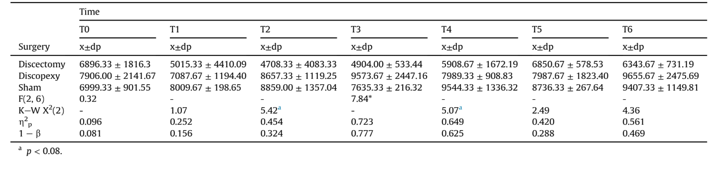
Deskriptive Basisstatistiken sind in Tabelle 1 dargestellt. Vier Ergebnisse wurden analysiert: (1) absolute Kaudauer; (2) Wiederkäuzeit pro Zyklus; (3) wiederkäuende Kinematik; und (4) wiederkäuende Fläche.
1) Absolute Kaudauer
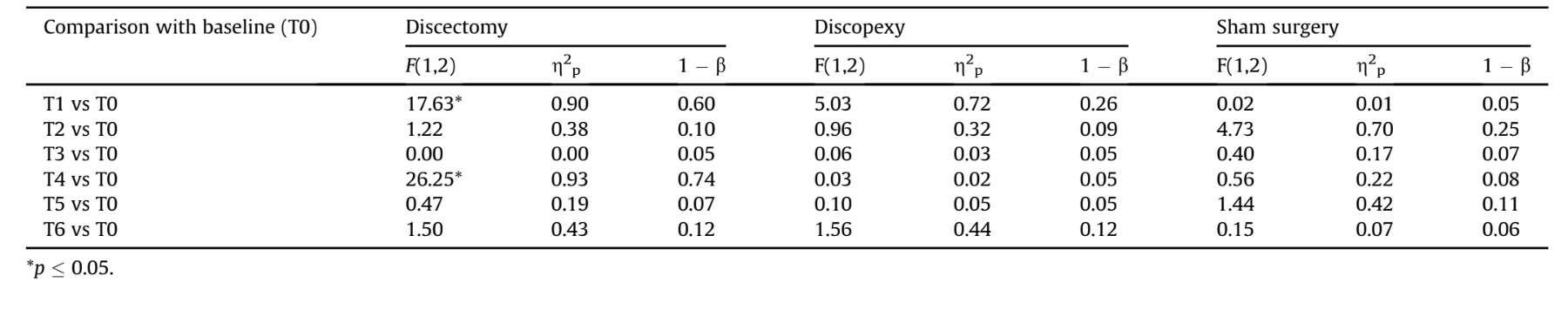
Querschnittsanalyse: Die Autoren verglichen die absoluten Kaudauern für die drei Gruppen jeden Monat nach der Operation (T1 bis T6). Eine einseitige ANOVA (oder das nicht-parametrische Äquivalent, der Kruskal-Wallis-Test) wurde durchgeführt, die signifikante Unterschiede zwischen den drei Gruppen nur in T1 zeigte - p = 0.03 (einseitig), Effektgröße von η2p = 0.736 (1-β) = 0.804 (Tabelle 2), aufgrund der höheren Werte für Discopexie im Vergleich zur Scheinoperation, wie durch einen Games-Howell Post-hoc-Test gezeigt (p = 0.028). Während der Basislinie und des verbleibenden Nachbeobachtungszeitraums (T2-T6) wurden keine statistisch signifikanten Unterschiede zwischen den Bedingungen Discektomie, Discopexie und Scheinoperation gefunden (p > 0.20).
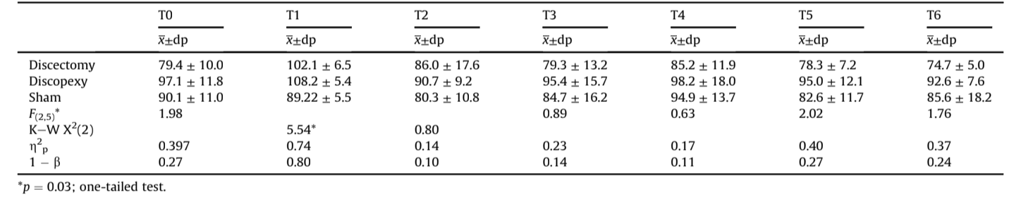
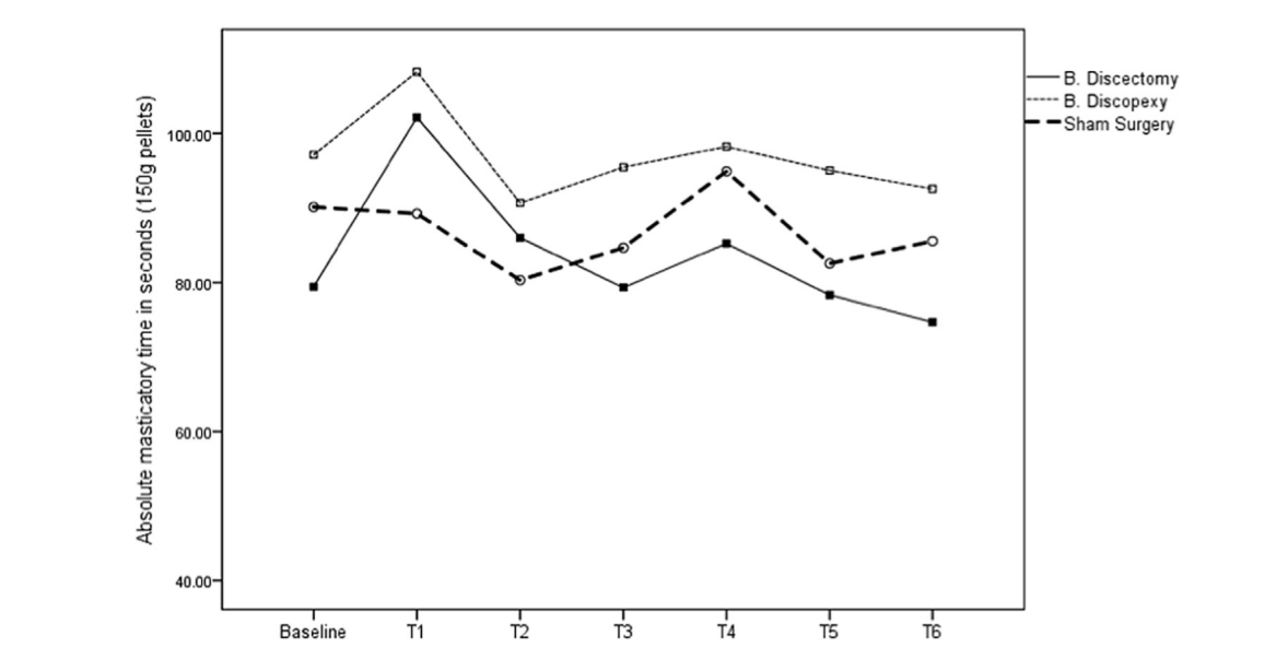
Langzeit Analyse: Eine einfaktorielle ANOVA mit wiederholten Messungen wurde durchgeführt, wobei als innerhalb der Probanden Effekte die Monate vor (T0) und nach der Operation (T1 bis T6) für die Bedingungen Diskektomie, Diskopexie und Scheinoperation betrachtet wurden. Signifikante Effekte über die Zeit wurden für die Diskektomie gefunden - F(6, 12) = 5.67, p = 0.005, η2p = 0.739 (1-β) = 0.947, jedoch nicht für Diskopexie und Scheinoperation - F(6, 12) = 2.65 und 1.59, p > 0.07, η2p = 0.570 und 0.443 (1-β) = 0.635 und 0.403, jeweils. In Anbetracht der Unterschiede im Verhältnis zur Basislinie (Tabelle 3) identifizierten die innerhalb der Probanden durchgeführten Kontraste einen statistisch signifikanten Anstieg nur für die Diskektomie zwischen T0 und T1 (Effektgröße von 90%; beobachtete Power von 0.60) und zwischen T0 und T4 (Effektgröße von 93%; beobachtete Power von 0.74). Für Diskopexie und Scheinoperation waren die Unterschiede im Verhältnis zur Basislinie trotz der Effektgrößen und unter Berücksichtigung der niedrigen beobachteten Power nicht statistisch signifikant. Abb. 4 zeigt die absolute Kaudauer für die Basislinie und von T1 bis T6.
2) Wiederkäuzeit pro Zyklus
Querschnittsanalyse: Die Ruminationzeit pro Zyklusrate variierte nicht zwischen den Gruppen sowohl im Pre-Test (T0) als auch zu allen Zeitpunkten des Post-Tests (p > 0.20), wie in Tabelle 4 gezeigt.
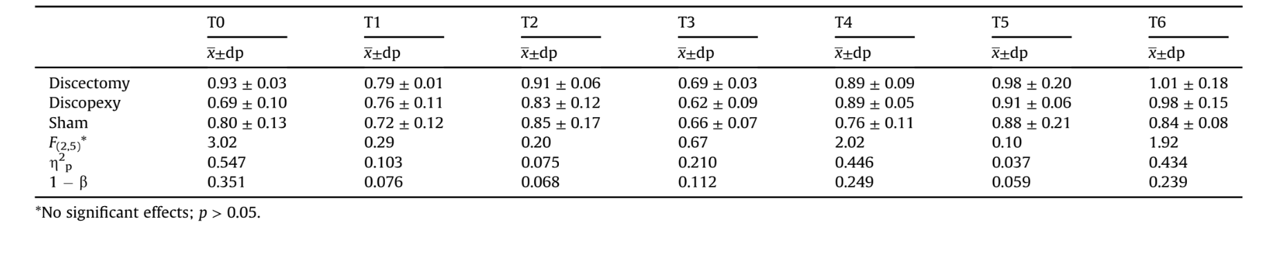
Langzeit Analyse: Eine einseitige ANOVA mit wiederholten Messungen wurde durchgeführt, wobei als innerhalb der Subjekte Effekte die Basislinie und die 6 Monate nach der Operation für Diskektomie, Diskopexie und Scheinoperation betrachtet wurden (siehe Tabelle 5).
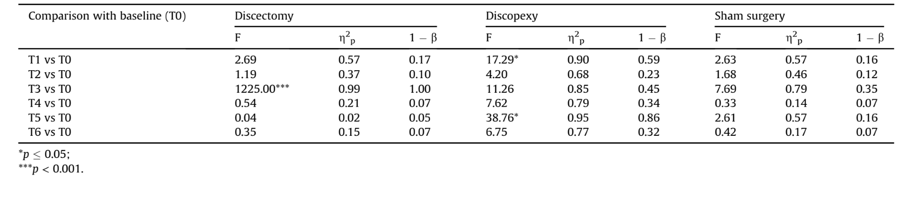
Eine signifikante Wirkung über die Zeit wurde für Discopexie und Scheinoperation gefunden - F(6, 6) = 6.87 und 4.11, p < 0.018, η2p = 0.773 und 0.673 (1-β) = 0.977 und 0.845, jeweils, aber nicht für Diskektomie - F(6, 6) = 2.70, p = 0.126, η2p = 0.730 (1-β) = 0.455.
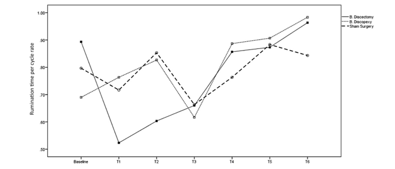
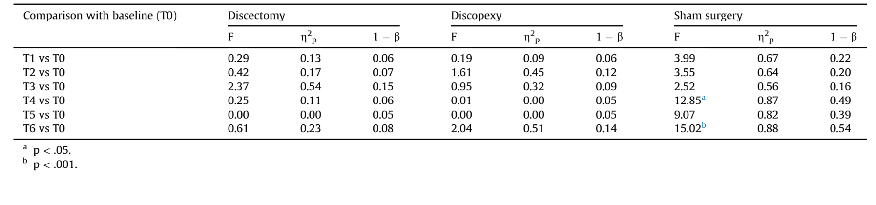
Der Vergleich der Wiederkäuzeit pro Zyklusrate zwischen der Basislinie und den Monaten nach der Operation identifizierte zwei Unterschiede für Discopexie, (T5 vs. T0) mit einer akzeptablen Power (Effektgröße von 95%). Für Diskektomie und Scheinoperation wurden keine signifikanten Unterschiede im Vergleich zur Basislinie gefunden. Abb. 5 zeigt die Wiederkäuzeit pro Zyklusrate in der Basislinie und von T1 bis T6. Wie zu sehen ist, wurden niedrigere Werte für die Zeiten T1, T2 und T3 erzielt, was darauf hindeutet, dass die Erholung des Schaf-TMJ bei T4 begann.
3) Ruminationskinematik und Fläche
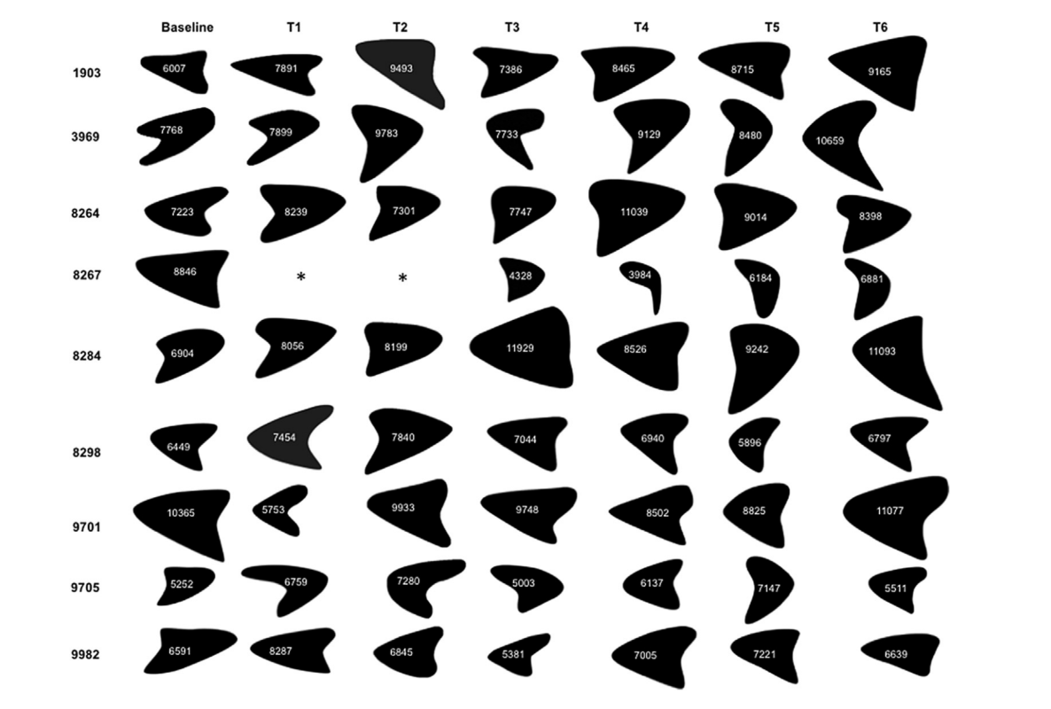
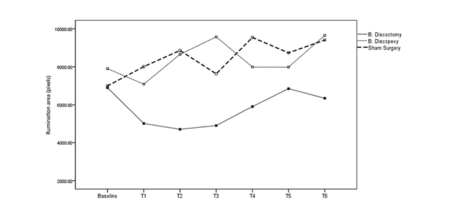
Beschreibende Ergebnisse zur Ruminationskinematik und zur durchschnittlichen Fläche der Ruminierung sind in Abb. 6 dargestellt.
Querschnittsanalyse: Die Rumination Bereiche variierten nur zwischen den Gruppen in T3 und T4. Für T3 identifizierte der Fisher LSD Post-hoc-Test eine signifikante Überlegenheit für den Discopexie-Bereich im Vergleich zum Diskektomie-Bereich (p = 0.008) (siehe Tabelle 6).
Langzeitanalyse: Eine einfaktorielle ANOVA mit wiederholten Messungen, mit Greenhouse-Geisser-Korrektur, die als innerhalb der Subjekte Effekte die Basislinie und die 6 Monate nach der Operation (T1 bis T6) für Diskektomie, Diskopexie und Scheinoperation betrachtet, zeigte keine statistisch signifikanten Unterschiede für die drei Bedingungen (p > 0,10). Die Unterschiede zwischen den Zeiten vor und nach dem Test waren ebenfalls nicht statistisch signifikant (p > 0,05), mit geringer Power, da (1-β) < 0,80, wie in Tabelle 7 zu sehen ist. Abb. 7 zeigt die Ruminationsbereiche für T0 (Vor-Test) und T1 bis T6 (Nach-Test) für Diskektomie, Diskopexie und Scheinoperation. Die Baseline-Ergebnisse sind für die drei experimentellen Bedingungen ähnlich. Nach der Operation waren die Ruminationsbereiche in der Diskektomie-Bedingung geringer, obwohl die Unterschiede nicht statistisch signifikant waren.
Unerwünschte Ereignisse
Es wurden keine schwerwiegenden unerwünschten Ereignisse gemeldet, abgesehen von einem Schaf in der Diskektomie-Gruppe, das in T1 und T2 das Wiederkäuen eingestellt hat, aber in T3 bis T6 zur normalen Funktion zurückkehrte.
Diskussion
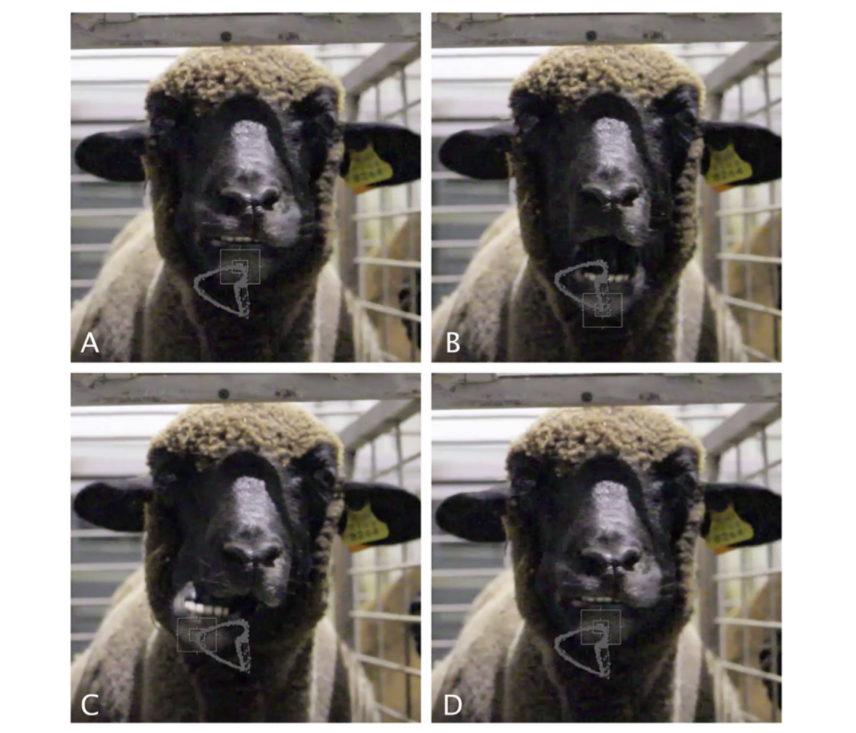
Das Hauptziel dieser Studie war es, die Auswirkungen verschiedener chirurgischer Eingriffe auf das Wiederkäuen und die Kaubewegungen von Schafen zu analysieren. Die vorgeschlagene Methodik hat sich als machbar und empfindlich gegenüber den Interventionen erwiesen. Homogene Bedingungen wurden in der Ausgangssituation erzielt und die Tiere verhielten sich natürlich vor der Kamera, was die Qualität der kinematischen Bewertungen gewährleistete (Abb. 3).
Die Messung der Kinematik wurde entwickelt, um das Verständnis der Auswirkungen von Kiefergelenkoperationen auf die Kieferbewegungen zu fördern. Theoretisch kann eine bilaterale Kiefergelenkoperation Veränderungen der Kieferbewegungen verursachen, aber diese Ergebnisse müssen quantifiziert werden.
Bezüglich der absoluten Kaudauer wurde erwartet, dass die Tiere nach einer bilateralen Diskektomie mehr Zeit benötigen, um die 150 g Pellets zu fressen (Ingawale und Goswami, 2009). Dementsprechend erhöhte die Diskektomiegruppe die Kaudauer um 28 % in T1. Dies könnte mit Schmerzen im Kiefergelenk zusammenhängen, was zu einer langsameren Nahrungsaufnahme führt. Am Ende der Studie waren diese Tiere in der Lage, sich auf Basiswerte (74,67 s) zurückzuziehen (Abb. 4). Wie bereits erwähnt, gibt es einen Mangel an Studien, die die Auswirkungen von Interventionen auf die Funktionalität des Kiefergelenks bewerten. Daher war es nicht möglich, dieses kaudynamische Ergebnis mit anderen Ergebnissen zu vergleichen. Obwohl es keine statistischen Unterschiede zwischen der Kaudauer vor und nach der Operation (d.h. T0 vs T1) gab, war der Unterschied spürbar. Nach T1 deutet die anschließende Rückkehr zu den Basiswerten darauf hin, dass Schafe die Fähigkeit hatten, sich an die induzierten Einschränkungen anzupassen, was die Bedeutung der Funktion über die Form hervorhebt (Poveda et al., 2007). Schafe haben, wie andere Tiere auch, die Fähigkeit, sich anzupassen, um zu überleben, selbst im Falle von größeren Eingriffen am Kiefergelenk, bei denen schwere Dysfunktionen zu katastrophalen Folgen für das Tier führen könnten.
Die Autoren sind sich einig, dass es interessant wäre, in Zukunft dieses Ergebnis über einen längeren Zeitraum zu analysieren.
Bezüglich der Wiederkäuzeit pro Zyklus wurden bemerkenswerte Ergebnisse erzielt. In der Diskektomie-Gruppe hörte ein Tier während T1 und T2 mit dem Wiederkäuen auf. Dies deutet auf die Notwendigkeit zukünftiger Untersuchungen in diesem Bereich hin, um beispielsweise zu verstehen, ob das Kiefergelenk einen wichtigeren Einfluss auf das Wiederkäuen als auf das Kauen haben könnte. Die Autoren glaubten zunächst, dass eine Ankylose nach bilateraler Diskektomie auftreten könnte, aber zu T3 wiederkäuten alle Tiere. Dieses Ergebnis deutet darauf hin, dass die Schafe trotz einer anfänglichen Verlangsamung im Zusammenhang mit der Nahrungsaufnahme, dem Wiederkäubereich und sogar einem Tier, das nicht wiederkäuen konnte, in der Lage waren, sich wieder anzupassen und zur normalen Partikelzerkleinerung zurückzukehren. Bei der Analyse von Abb. 5 ist auffällig, dass alle Gruppen die Wiederkäuzeit pro Zyklus in T3 reduzierten, ohne die Ursache für ein Ereignis zu kennen, das zu diesem Ergebnis führte. In T4eT6 nahmen die Schafe jedoch wieder die erwarteten Werte an. Die Tiere aus den Gruppen Diskektomie und Diskopexie benötigten in T5 und T6 mehr Zeit, um einen Wiederkäuzug zu erreichen, was auf einen weniger effektiven Wiederkauprozess hindeutet.
Bezüglich des Ruminationsbereichs ist auffällig, dass ein schnellerer Ruminationszyklus durch einen kleineren Ruminationsbereich erzielt wird. Ein weiteres interessantes Detail ist, dass in T3 und T4 eine Normalisierung der Ruminationskinematik für die Diskektomie-Gruppe beobachtet wurde. Dieses Ergebnis deutet darauf hin, dass eine Umgestaltung und Anpassung 3-4 Monate nach der chirurgischen Intervention am Kiefergelenk erfolgt. Obwohl die Ruminationsbereiche in der Diskektomie-Gruppe nach der Operation reduziert wurden, waren die Unterschiede statistisch nicht signifikant.
Die Bewertung der Trajektorie und des Ruminationsbereichs war interessant, da es möglich war, ein Muster zu identifizieren. Jedes Tier zeigte eine bevorzugte Seite für die Ruminierung, wechselte jedoch unabhängig von der Intervention die Seiten. Jedes Tier zeigte eine dreieckige Trajektorie, ähnlich den Kieferbewegungen, die bei narkotisierten Kaninchen demonstriert wurden (Hidaka et al., 1997).
Weitere Forschungen sollten in der Lage sein, mögliche Zusammenhänge zwischen diesen Ergebnissen und histologischen, bildgebenden und Gewichtsergebnissen zu untersuchen (Zhao et al., 2010).
Schlussfolgerungen
Die Autoren sind sich keiner vorherigen randomisierten, verblindeten, präklinischen Studien im Bereich des Kiefergelenks bewusst, die den ARRIVE-Richtlinien folgen. Mit schwarzen Merinoschafen, unter Berücksichtigung von Alter und Geschlecht, einem öffentlich verfügbaren Protokoll, einer Schein-Kontrollgruppe und einem bilateralen Ansatz beabsichtigten wir, mögliche Verzerrungen zu minimieren. Der bilaterale Ansatz vermied auch negative Auswirkungen des nicht operierten kontralateralen Gelenks, wie sie bei einseitigen Verfahren berichtet wurden (Dimitroulis und Slavin, 2006). Die vorgeschlagenen Basisergebnisse waren homogen und die Schein-Kontrollgruppe funktionierte effektiv.
Der erste Monat nach der Intervention scheint der kritische Zeitraum in Bezug auf kinematische Veränderungen zu sein, mit Modifikationen, die sich auf die absolute Kaudauer, die Wiederkäuzeit pro Zyklus und die Wiederkäufläche beziehen, sowohl in den Gruppen der Diskektomie als auch der Diskopexie. Nach 1 Monat scheint die bilaterale Diskopexie des Kiefergelenks keinen wichtigen kinematischen Einfluss bei schwarzen Merinoschafen zu haben. Die bilaterale Diskektomie des Kiefergelenks scheint jedoch einen signifikanten Einfluss zu haben, hauptsächlich in T1 und T2, aber von T3 bis T6 wird eine Normalisierung der Ergebnisse beobachtet.
Die Autoren sind sich einig, dass das rigorose Studiendesign, das Tiermodell und die bilaterale Intervention die Hauptvorteile dieser Forschung waren. Die Einschränkungen waren hauptsächlich auf die kleine Stichprobengröße zurückzuführen, daher sollte die weitere Forschung auf größere Stichproben abzielen. Die Einführung der kinematischen Bewertung hebt die Bedeutung der Kinematik hervor.
David Faustino Ângelo, Florencio Monje Gil, Raúl González-García, Lisete Mónico, Rita Sousa, Lia Neto, Inês Caldeira, Carla Moura, Luís Carlos Francisco, David Sanz, Nuno Alves, Francisco Salvado, Pedro Morouço
Literaturverzeichnis
- Ângelo DF, Monje FG, Gonzalez-Garcia R, Little CB, Monico L, Pinho M, et al: Bioengineered temporomandibular joint disk implants: Studienprotokoll für eine zweiphasige explorative randomisierte präklinische Pilotstudie an 18 schwarzen Merino-Schafen (TEMPOJIMS). JMIR Res Protoc 6: e37, 2017
- Angelo DF, Morouco P, Alves N, Viana T, Santos F, Gonzalez R, et al: Auswahl von Schafen (Ovis aries) als Tiermodell für die Forschung zum Kiefergelenk: morphologische, histologische und biomechanische Charakterisierung der Gelenkscheibe. Morphologie 100: 223-233, 2016
- Bjørnland T, Haanaes HR: Discektomie des Kiefergelenks: eine experimentelle Studie an Affen. J Craniomaxillofac Surg 27: 113-116, 1999
- Bjørnland T, Larheim TA: Discektomie des Kiefergelenks: 3-Jahres-Nachuntersuchung als Prädiktor für das 10-Jahres-Ergebnis. J Oral Maxillofac Surg 61: 55-60, 2003
- Cohen WA, Servais JM, Polur I, Li Y, Xu L: Degeneration des Gelenkknorpels im kontralateralen nicht-chirurgischen Kiefergelenk bei Mäusen mit einer einseitigen partiellen Discektomie. J Oral Pathol Med 43: 162-165, 2014
- Dimitroulis G, Slavin J: Die Auswirkungen der einseitigen Discektomie und Kondylektomie auf das kontralaterale intakte Kaninchen-Kranio-Mandibular-Gelenk. J Oral Maxillofac Surg 64: 1261-1266, 2006
- Eriksson L, Westesson P-L: Discektomie als effektive Behandlung für schmerzhafte interne Störungen des Kiefergelenks: eine 5-jährige klinische und radiografische Nachuntersuchung. J Oral Maxillofac Surg 59: 750-758, 2001
- Hidaka O, Morimoto T, Masuda Y, Kato T, Matsuo R, Inoue T, et al: Regulierung der Kaumuskraft während kortikal induzierter rhythmischer Kieferbewegungen im narkotisierten Kaninchen. J Neurophysiol 77: 3168-3179, 1997
- Ingawale S, Goswami T: Kiefergelenk: Störungen, Behandlungen und Biomechanik. Ann Biomed Eng 37: 976e996, 2009
- Kilkenny C, Parsons N, Kadyszewski E, Festing MFW, Cuthill IC, Fry D, et al: Umfrage zur Qualität des Studiendesigns, der statistischen Analyse und der Berichterstattung von Forschung mit Tieren. PLoS One 4: e7824, 2009
- Mazzonetto R, Spagnoli DB: Langzeitbewertung der arthroskopischen Discektomie des Kiefergelenks unter Verwendung des Holmium-YAG-Lasers. J Oral Maxillofac Surg 59: 1018-1023, 2001
- Nyberg J, Adell R, Svensson B: Discektomie des Kiefergelenks zur Behandlung einseitiger interner Störungen – eine 5-Jahres-Nachuntersuchung. Int J Oral Maxillofac Surg 33: 8-12, 2004
- Ogi N, Kurita K, Ishimaru J, Goss AN: Kurzzeitige Wirkung der tiefgefrorenen, gelagerten aurikulären Knorpel-Allograft-Reparatur auf das osteoarthritische Kiefergelenk des Schafes. Int J Oral Maxillofac Surg 28: 393-397, 1999
- Pearce G: Veränderungen der Partikelgröße im Retikulum-Rumen von Schafen. Aust J Agric Res 18: 119, 1967
- Poveda RR, Bagan JV, Diaz Fernandez JM, Hernandez Bazan S, Jimenez SY: Übersicht über die Pathologie des Kiefergelenks. Teil I: Klassifikation, Epidemiologie und Risikofaktoren. Med Oral Patol Oral Cir Bucal 12: e292-e298, 2007
- Sato S, Goto S, Koeda S, Motegi K: Veränderungen des elastischen Faser-Netzwerks des Kiefergelenks des Kaninchens nach Discektomie. J Oral Rehabil 29: 847-852, 2002
- Sharma R, Sinha R, Menon PS: Meniskopexie bei interner Störung des Kiefergelenks. J Maxillofac Oral Surg 13: 261-265, 2010
- Takaku S, Sano T, Yoshida M: Langzeit-Magnetresonanztomographie nach Discektomie des Kiefergelenks ohne Ersatz. J Oral Maxillofac Surg 58: 739-745, 2000
- Tong AC, Tideman H: Eine vergleichende Studie zu Meniskektomie und autologer Transplantation des Gelenkknorpels des Rhesusaffen-Kiefergelenks: Teil I. Int J Oral Maxillofac Surg 29: 140-145, 2000
- Trumpy IG, Lyberg T: Chirurgische Behandlung von internen Störungen des Kiefergelenks: Langzeitbewertung von drei Techniken. J Oral Maxillofac Surg 53: 740-747, 1995
- Van der Worp HB, Howells DW, Sena ES, Porritt MJ, Rewell S, O'Collins V, et al: Können Tiermodelle von Krankheiten zuverlässig Informationen für menschliche Studien liefern? PLoS Med 7: e1000245, 2010
- Zhao C, Kurita H, Kurashina K, Hosoya A, Arai Y, Nakamura H: Reaktion des Kiefergelenks auf die Mandibularabweichung bei Kaninchen, erkannt durch 3D-Mikro-CT-Bildgebung. Arch Oral Biol 55: 929-937, 2010